or flies, the larva develops into adult worm (4-10mm long) which after mating produce many microfilaria
(170-320mm long ) that migrate in blood or skin typically, the disease is due to host immune response to the worm (particularly
dying worms).
Wuchereria Bancrofti (in about 90% of cases), Brugia Malayi (9%) and Brugia Timori (1%) are the filarial worms responsible for
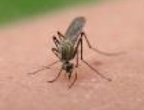
lymphatic filariasis. All are transmitted by either of Anopheles (in rural areas), Culex (urban and semi urban areas) or Aedes
(endemic islands in the pacific) mosquitoes.
BURDEN OF LYPHATIC FILARIASIS
More than 1.3 billion people in 81 countries world wide are threatened by lymphatic filariasis.
Over 120 million people are currently infected, with about 40 million disfigured and incapacitated by the disease (25
million men with genital disease, and over 15 milllion people with lymphedema)
PATHOGENESIS (LIFE CYCLE OF THE FILARIAL WORM)
When a mosquito with infective stage larvae bites a person, the parasites are deposited on the skin from where they enter the
body. The larvae then migrate to the lymphatic vessels where they develop into adult worm forming ‘nests’ in the human
lymphatic system. The adult worm release toxins which cause lymphangiectasia (dilatation of the lymphatic vessel) leading to
lymphatic dysfunction and the chronic clinical manifestation of the disease lymphedema due to inability of the lymph vessels to
drain fluid adequately.
Death of the adult worms (which can live up to 10-15 years) result in acute filarial lymphangitis. Release of lipopolysaccharide
from these bacteria contributes to inflammation.
The adult female worm produces many microfilariae which circulate in large number in the peripheral blood, usually at night.
The life cycle is complete when the vector (mosquito) takes up microfilariae during a blood meal.
CLINICAL PRESENTATION
The disease involves asymptomatic, acute and chronic conditions:
Asymptomatic condition make up the majority of the infections, showing no external sign of infection these
asymptomatic infection still cause damage to the lymphatic system the kidneys and immune system.
Acute episodes of local inflammation involving skin, lymph nodes and lymphatic vessels often accompany the chronic
manifestation. They include:
– High fever
– Red hot swollen leg
– Chills
– vomiting
– Headache
Most of these are caused by body’s immune responses of the parasite.
Lymphedema (tissue swelling due to fluid accumulation) affecting the limbs, genital organ and breast
Elephantiasis (thickened and darkened skin tissue) of the limbs and genitals.
Hydrocele (fluid accumulation) affecting the scrotum, penis, etc.
Tropical pulmonary eosinophilia which is a complication due to destroyed trapped microfilaria in pulmonary capillaries.
TREATMENT
The following treatment modalities are employed
1. Medical treatment
– Albendazole (400mg) plus ivermectin (150-200mg/kg)
For onchocerciasis endemic areas eg the tropics.
– Albendazole plus diethylcarbamazine (DEC) (6mg/kg) for onchocerciasis non endemic areas.
– Antibiotic, doxycycline (200mg) for 8 weeks kills the symbiotic bacteria, wolbacha, leading to the death of the worm.
2. Surgical treatment
This may be helpful for issues related to scrotal elephantiasis and hydrocele but research has shown that medical management
is better.
3. Rigorous daily cleaning of the affected areas of the body limits symptoms of lymphatic filariasis.
PREVENTION
a. Control of mosquito with nets, sprays, etc in endemic zone
b. Mass drug administration (albendazole + ivermectin /DEC)
REFERENCES
1. http://www.who.int/mediacentre/factsheet.
2. Davidson’s principles and practice of medicine
3. en.m. Wikipedia.drug/ wiki/elephantiasis
4. Elephantiasis free yellow. Com/
5. www.hxbenfit.com/ elephantiasis.html.
6. www.mamashealth.com/parinfect/elep.asp